
Through ‘Quantitative Feulgen Micro-spectrophotometry DNA Stain method’, they observed that if the light is being passed through interphase, the cot values (described below) were Said that DNA replication occurs in Prophase.

He named the thread-like structures ‘chromosomes’. Observed that the thread-like structure took a certain stain. Observed, along with nucleus, some thread-like structures were also divided equally and name the incident ‘Mitosis’ (mito =thread, sis= division). German biologist and a founder of cytogenetics Here, ‘karyo’ = nucleus & ‘kinesis’ = division. Both prokaryotic & eukaryotic organisms grow up and reproduce by this process.Īlso, 3 types of cell divisions occur in living organisms:įirst gave the word ‘Karyokinesis’. However, it occurs as a small part of a cell cycle. Cell DivisionĬell division is the basic process by which a parent cell divides into two or more daughter cells. Multi-cellular organisms grow up and reproduce by dividing their cells. For instance, higher plants, animals, human beings, etc. Thus, these are called multi-cellular organisms. In contrast, there are also many other organisms whose bodies are made up of two or more cells. Some organisms have only one cell during their whole lifespan by which they carry out all the physiological processes they require to survive. Prophase is generally considered to be over when the chromosomes are fully condensed, clear, and the nuclear membrane is gone or almost gone.All living things, as well as human beings, are made up of cells.

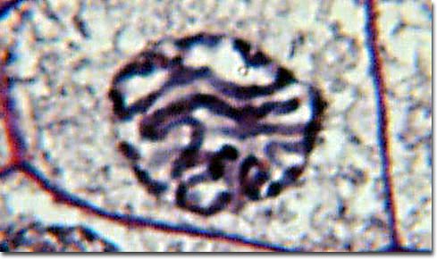
The centrioles, and asters, are at opposite ends of the cell and the thin protein spindle fibers are reaching out and attaching to the centromeres of each chromosome from opposite directions. The chromosomes are very distinct, easy to recognize and have clear "arms" composed of the two parts of the sister chromatids. Late prophase - the nuclear membrane and the nucleolus finally vanishes completely. In animal cells a system of thin protein fibers begins to radiate out from the centrioles forming a pattern in the cytoplasm of the cell that looks like a star or aster. The pairs of centrioles continue to move around the almost vanished nucleus to opposite sides of the cell. The location of the centromere along the length of the chromosome and chromatids is a distinctive characteristic of many chromosomes, and can sometimes be used (with other factors) to identify them. Each chromosome consists of two identical halves called chromatids, that are connected and held together by a constriction called a centromere. Mid-prophase - the chromatin threads are now condensed enough to be distinguished as individual chromosomes. In animal cells (mostly), a double pair of short, rod-like structures called centrioles, appear, separate and begin to move to opposite sides of the cell, outside the vanishing nucleus.

The nucleolus also becomes indistinct and begins to vanish. It is usually not possible to follow individual threads, but the condensation of the material into individual units is becoming obvious. This is generally taken as the beginning of prophaseĮarly prophase - the nuclear membrane becomes more and more indistinct and the chromatin fibers become more and more packaged and condensed. The molecules of DNA become associated with more and more histone proteins and package themselves into higher and higher degrees of structure.
#PROPHASE UNDER MICROSCOPE SERIES#
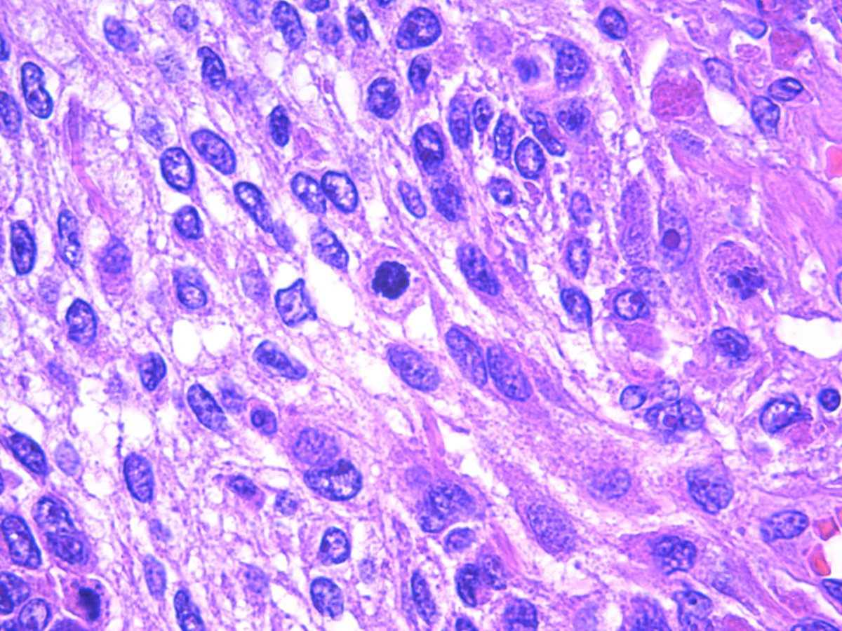
ProphaseĪs the cell moves out of G 2 and into M the granular nature of the nucleus begins to change and condense into a series of fine threads. Nuclear division or karyokinesis is a continuous process, however, and there are no artificial divisions in actively growing cells. Early microscopists found it convenient to subdivide the nuclear division of cells into stages that were easily seen under the microscope using colored dyes that stained the chromosomes and some of the other participants. With the right techniques, the next stage in the cell cycle, mitosis (M), can be observed using a good light microscope. The three main phases of a single cell cycle are: interphase, nuclear division and cytoplasmic division.


 0 kommentar(er)
